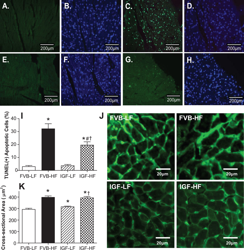
Fig. 4.
Effect of IGF-1 overexpression on myocardial apoptosis and hypertrophy following LF or HF feeding using TUNEL and FITC-conjugated Lectin staining, respectively. All nuclei were stained with DAPI (blue) in panels B (FVB-LF), D (FVB-HF), F (IGF-LF) and H (IGF-HF). TUNEL-positive nuclei were visualized with fluorescein (green) in panels A (FVB-LF), C (FVB-HF), E (IGF-LF) and G (IGF-HF). Original magnification = 400×. Quantified data are shown in panel I; J: FITC-conjugated Lectin immunostaining depicting transverse sections of left ventricular myocardium (×400); and K: Quantitative analysis of cardiomyocyte cross-sectional area. Mean ± SEM, n = 15 and 10 fields from 3 mice per group for panel I and K, respectively, *p < 0.05 vs. FVB-LF group; #p < 0.05 vs. FVB-HF group, †p < 0.05 vs. FVB-LF group.

