
Figure 2. Embryonic expression of Ntla-dependent genes.
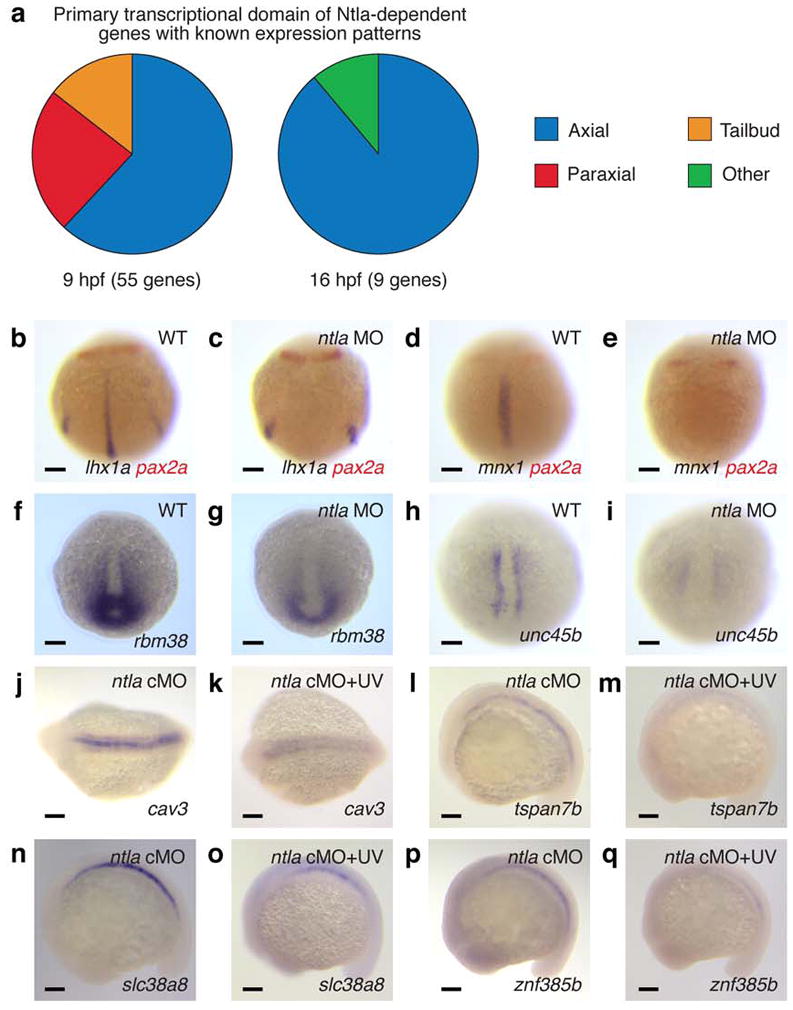
a, Tissue localization of Ntla-dependent genes that have known expression patterns and are transcribed during gastrulation (9 hpf) or somitogenesis (16 hpf). b-i, Confirmation of selected microarray hits by in situ hybridization. 10-hpf wildtype and ntla MO-injected embryos stained for candidate Ntla targets expressed during gastrulation are shown, with co-labeling of pax2a transcripts to determine embryo orientation if necessary. j-q, Analogous studies of candidate Ntla targets expressed during somitogenesis. 16-hpf ntla cMO-injected embryos that were either cultured in the dark or globally UV irradiated at 12 hpf are shown. Embryo orientations: b-e and h-i, dorsal view and anterior up; f-g, dorsal posterior view and dorsal up; j-k, dorsal view and anterior left; l-q, lateral view and anterior left. Scale bars: 100 μm.