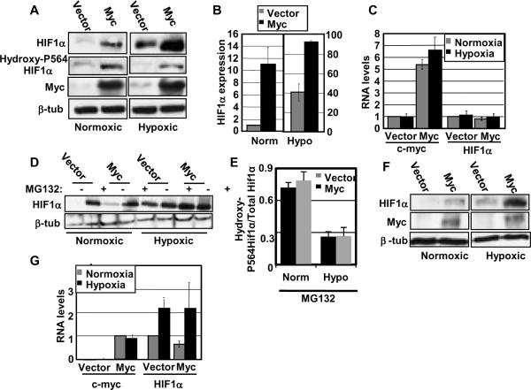
Figure 1. Myc post-transcriptionally enhances accumulation of HIF1α under both normoxia and hypoxia.
A) Western blot analyses of IMECs were performed with equivalent amounts of protein using anti-HIF1α antibody, anti-hydroxyl P564-specific HIF1α antibody (OH-P564), anti-c-Myc antibody, and anti-β-tubulin antibody. Hypoxic cell extracts were made after incubating cells for two hours in a hypoxia chamber with 1% oxygen. Normoxic HIF1α blots are shown at a longer exposure than the hypoxic HIF1α blots. Norm= Normoxia, Hypo= Hypoxia. B) Average HIF1α protein expression, normalized to β-tubulin, was quantitated using three independent cell extract sets. Expression levels are normalized to normoxic vector. C) Real-time RT-PCR was performed using RNA harvested from IMECs and primers specific to HIF1A, c-myc, and GAPDH in triplicate. Average c-myc and HIF1A RNA levels are normalized to GAPDH and then normalized to their respective normoxic vectors. D) IMECs were treated with proteasome inhibitor MG132 under normoxic and hypoxic conditions. Western blot analyses were performed with equivalent amounts of protein using anti-HIF1α and anti-β-tubulin antibodies. E) After treatment with MG132, average hydroxy-P564 HIF1α protein expression was determined in comparison to total HIF1α protein expression under normoxia and hypoxia. F) Western blot analyses of myc−/− rat fibroblasts (vector or Myc-reconstituted) were performed as described above. G) Real-time RT-PCR was performed using RNA harvested from myc−/− rat fibroblasts and primers specific to HIF1A, c-myc, and GAPDH in triplicate. Average c-myc and HIF1A RNA levels are normalized to GAPDH. HIF1A is normalized to its normoxic vector. c-myc is normalized to normoxic myc−/−(Myc) since vector cells express no c-myc RNA. Error bars reflect the standard deviation.

