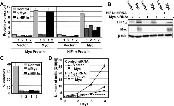
Figure 7. HIF1α expression is affected by Myc levels and is necessary for anchorage-independent and log phase growth.
A, B) IMECs were transfected with non-targeting siRNA or siRNAs (1 or 2) targeting HIF1A or c-myc. Cell extracts were made 48 hrs post-transfection. Western blot analyses were performed with equivalent amounts of cell extracts using anti-HIF1α, anti-c-Myc, and anti-β-tubulin antibodies. Average Myc and HIF1α protein expression, normalized to β-tubulin, were quantitated using three independent cell extract sets. Expression levels for Myc and HIF1α protein were normalized to their respective vectors. C) IMECs transfected with non-targeting siRNA or siRNAs (1 or 2) targeting HIF1A or c-myc were plated in soft agar 24 hours post-transfection. The number of colonies per 100 plated Myc-overexpressing cells were counted and measured after 10 days in two independent experiments. Vector cells do not grow in soft agar and are therefore not included in the analysis. D) Cell proliferation rates were measured by counting equivalently plated cells that had been transfected at Day 1 with either HIF1A or non-targeting siRNA under normoxia. The graph shows the mean cell number for each day for duplicate experiments. Error bars reflect the standard deviation. Similar results were found for independent siRNAs.

