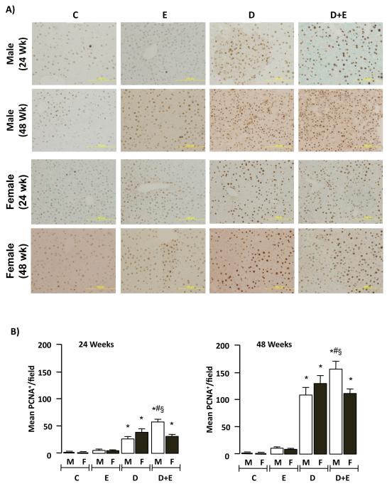
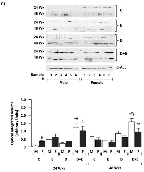
Figure 4. Ethanol feeding promotes hepatic tumorigenesis preferentially in male mice.
A) Representative proliferating cell nuclear antigen (PCNA) immunohistochmistry of hepatic sections (x200 magnification) from control (C) mice, or mice maintained on ethanol (E), DEN-initiation (D), or DEN and ethanol (D+E) at 24 or 48 weeks. B) Number of PCNA positive cells per microscopic field (x200 magnification) were measured in representative sections (2 lobes/mouse, 5 fields/lobe) from individual male and female mice on different treatment regimes (24 and 48 weeks) and mean values (± SEM) were calculated. *p<0.05 versus respective male or female E, #p<0.05 D+E versus respective male or female D mice, §p<0.05 male versus female within same treatment group/time points. Minimum n = 8 independent mice/group. C) Representative immunoblots of cyclin D1 expression in hepatic tissue lysates from male and female mice on different treatment regimes at 24 and 48 weeks (upper panel). Optical integrated volume was calculated, corrected for sample loading (stripping-re-probing membranes with anti-β-actin (β-Act)) and expressed as mean values (arbitrary units) ± SEM (lower panel). *p<0.05 versus respective male or female E, #p<0.05 D+E versus respective male or female D mice, §p<0.05 male versus female within same treatment group/time points. Minimum n = 6 independent samples per group.


