
Figure 1.
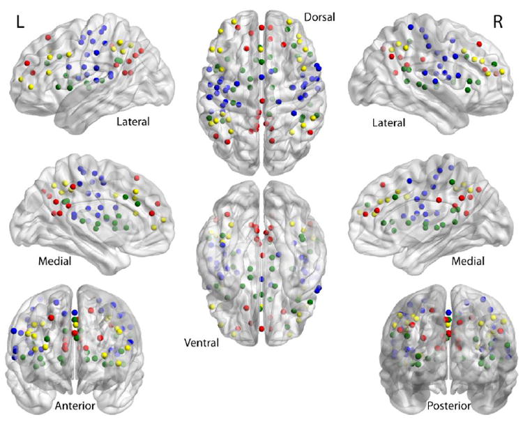
Shown are the 100 seed-regions used in this study taken from Dosenbach et al. (2010). Fronto-parietal network is in yellow; sensorimotor in blue; default in red; cingulo-opercular in green.
Official websites use .gov
A
.gov website belongs to an official
government organization in the United States.
Secure .gov websites use HTTPS
A lock (
) or https:// means you've safely
connected to the .gov website. Share sensitive
information only on official, secure websites.
Shown are the 100 seed-regions used in this study taken from Dosenbach et al. (2010). Fronto-parietal network is in yellow; sensorimotor in blue; default in red; cingulo-opercular in green.