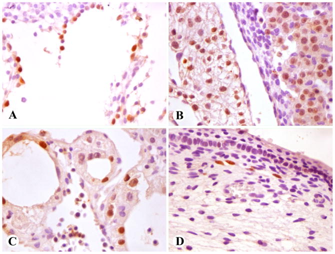
Fig. 1.
Prox1-positive developing tissues (nuclear positivity). A–C: Early first trimester embryo. D. Late first trimester fetus. A. A large vessel with flaccid contours, consistent with thoracic duct. B. Cardiac myocytes (left) and hepatocytes (right). C. Yolk sac epithelia. D. Subdermal lymphatic vessels in a limb.

