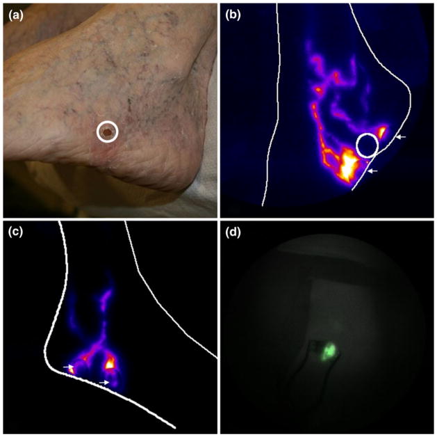
FIGURE 7.
(a) Color image of the tumor site (post biopsy) on the outer left foot of a 60 year old female with melanoma and NIRF images of (b) the tortuous lymphatics on the diseased foot, (c) the corresponding lymphatics on the contralateral limb, and (d) the SLN in the inguinal basin as seen during surgery. Circle is biopsy site, arrows show ICG injection sites. Images are displayed in pseudo color.

