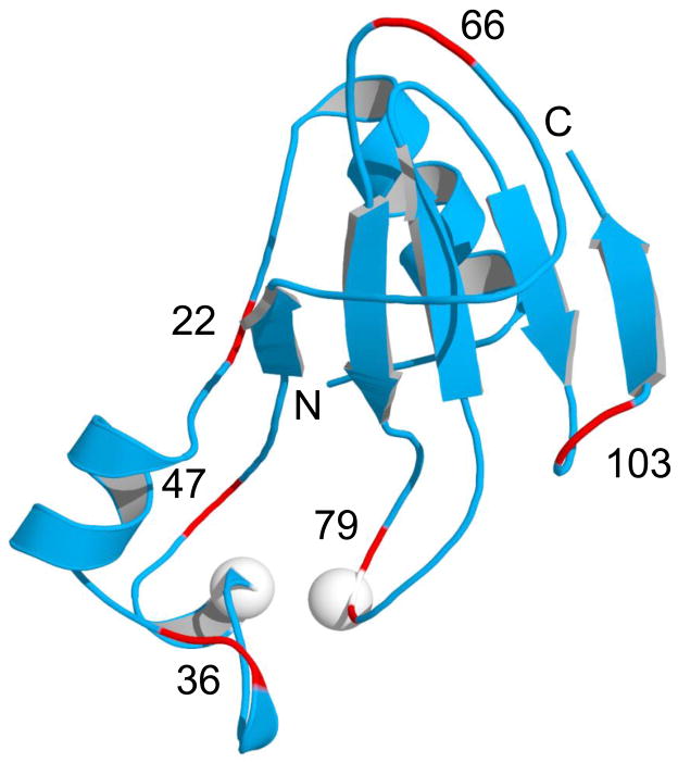
Figure 2.
Structure of WT Bn (PDB 1A2P) showing surface loops (red) where Ub is inserted. The Bn-Ub fusion proteins constructed in this study (BU22, BU36, BU47, BU66, BU79, and BU103) are named according to the Bn residue numbers indicated in the figure. White spheres denote positions of Cys residues in the BU66 (A43C+S80C) double mutant.
BU genes were created by inserting the Ub gene into the Bn gene at the positions indicated, following the procedure of Geiser et al. (24). The genes were fused using nucleotides encoding Gly-Gly and Gly as the first and second linkers, respectively, and the Ub gene lacked a codon for the N-terminal Met. BU proteins were purified as described (17).

