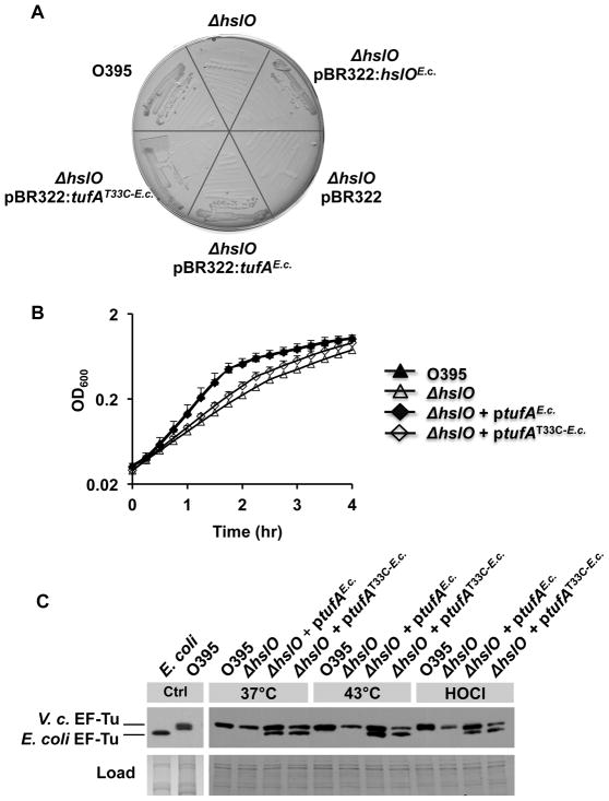
Fig. 5. E. coli EF-Tu compensates for lack of Hsp33 by protecting V. cholerae EF-Tu against oxidative protein degradation.
A. V. cholerae O395 wild type, O395 ΔhslO mutant, or O395 ΔhslO expressing the empty pBR322 vector, E. coli Hsp33, E. coli EF-Tu,or the E. coli EF-TuT33C variant were cultivated on MacConkey plates for 24 h at 43°C.
B. V. cholerae O395 wild type (black triangles), O395 ΔhslO mutant (white triangles), and O395 ΔhslO expressing either E. coli EF-Tu(black diamonds) or the E. coli EF-TuT33C variant (white diamonds)were cultivated in LB medium at 43°C aerobically. Bacterial growth was monitored by optical density measurements at 600 nm.
C. To analyze the steady state levels of EF-Tu in these bacterial strains, the different strains were cultivated in LB medium at either 37°C or 43°C until mid-log phase was reached. To evaluate the effects of HOCl on cellular EF-Tu levels, the 37°C cultures were split and either left untreated or incubated with 3 mM HOCl for 20 min. Cell aliquots were taken and the proteins were separated by SDS-PAGE. EF-Tu was visualized by Western blot using antibodies against E. coli EF-Tu.

