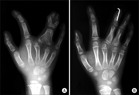
Fig. 16.
Case #14, roentgenogram at the age of six. (A) The united distal phalanges were separated and the space between these phalanges was filled with free fat graft at the age of three, but there was no recurrence of the bone union. (B) This was a roentgenogram 7 weeks after the syndactyly release of the middle and ring fingers. The united proximal phalanges were separated and the space between these phalanges was filled with free fat graft. The distal interphalangeal joint of the middle finger was unstable and it was fixed with a Kirschner wire for 8 weeks.

