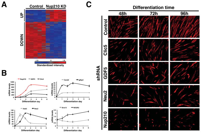
Figure 4. Nup210 regulates gene expression in post-mitotic myotubes.
(A) C2C12 cells were infected with lentivirus carrying control or Nup210 shRNAs at 36 hours after differentiation and whole genome expression was analyzed by microarray. Heat map shows the microarray expression profile of altered expression in Nup210 knock-down myotubes. See also Table 1 and S1. (B) The expression of Nup210, GDF5, Clic5, Asb2, Neu2, Cand2, Igfbp4, Stra13 and NDRG2 during myoblast differentiation was analyzed by qPCR. mRNA levels in differentiating cells were normalized to the levels of dividing cells. Values represent average ± SD of 3 different independent experiments. (C) C2C12 myoblasts were infected with lentivirus carrying control, Clic5, GDF5, Neu2 or Nup210 shRNAs and induced to differentiate. Immunofluorescence against MHC was performed at 48, 72, and 96h post differentiation. See also Figure S5.

