Fig. 1.
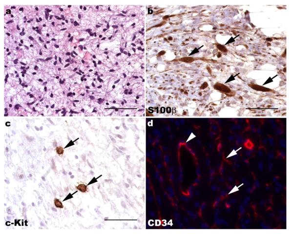
Neurofibromas are composed of a complex mixture of cell types. (a) Hematoxylin and eosin stained section of a plexiform neurofibroma demonstrating the bland spindle cells set against a myxoid background that are typically seen in these lesions. (b) Immunohistochemistry for the Schwann cell marker S100β highlights several immunoreactive elements within this plexiform neurofibroma (arrows). Note that numerous S100β-negative cells are also present. (c) Immunoreactivity for c-Kit (CD117; arrows) is evident in numerous mast cells in neurofibromas. (d) CD34 immunoreactivity (red) is present within both vasculature (arrowhead) and a dendritic population (arrows) in neurofibromas. The density of CD34-immunoreactive dendritic cells is variable in most neurofibromas; this region is particularly abundant in these cells. a-c, 40x (scale bars, 50 μm); d, 60x.

