
Figure 5. Screening of HePTP inhibitors in human T cells.
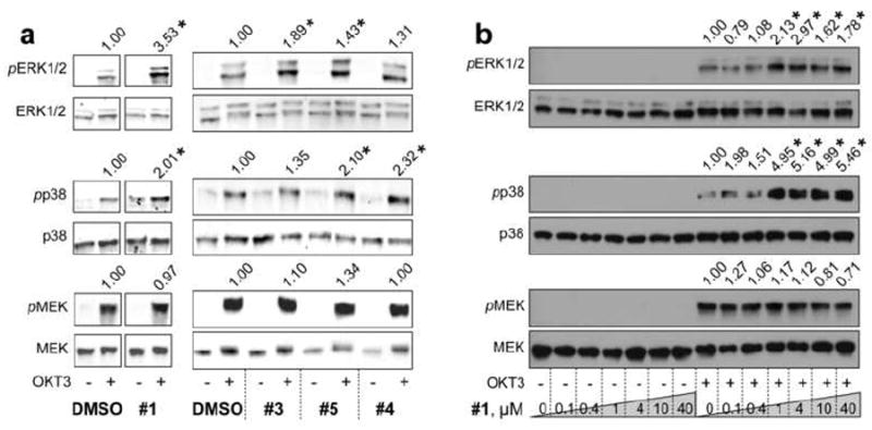
(a) Probing ERK1/2 and p38 phosphorylation in Jurkat TAg T cells in the presence of vehicle (DMSO, 0.2%) or compound 1, 3, 4, or 5 (40 μM), using phospho-specific antibodies against pT202/Y204-ERK1/2 (pERK1/2) and pT180/Y182-p38 (pp38). An anti-phospho-MEK (pMEK) blot was included as a specificity control. (b) Dose-response of compound 1. Cell lysates were probed as in (a). Phospho-protein bands were normalized to total protein levels. Statistical significance was determined by Student’s t-test (*p<0.05). Representative blots are shown from 3 independent experiments.