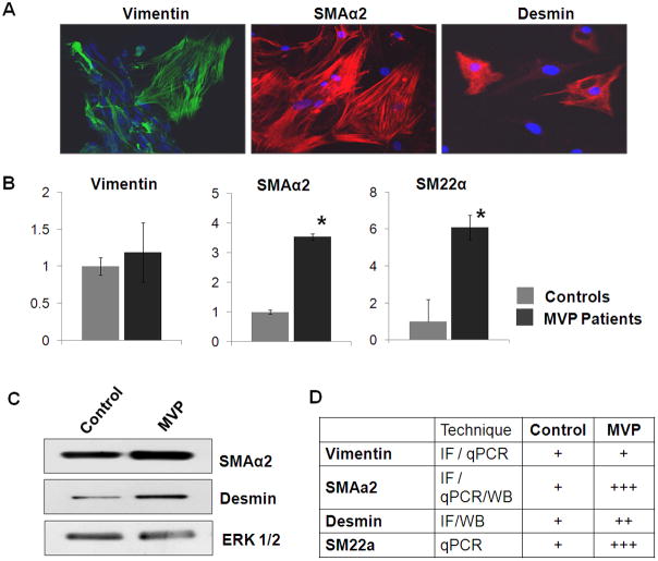
Figure 5. Characterization of Mitral VICs.
[A] Immunofluorescence staining for Vimentin, SMAα2, and Desmin in the isolated VICs. Magnification: 40X, Scale: 50μm. [B] Relative abundance of transcripts for Vimentin, SMAα2 and SM22α in MVP-derived VICs, using qPCR. (* denotes p<0.05). [C] Western blots showing expression of SMAα2, desmin and ERK1/2 (loading control) [D] Schematic representation for expression of VIC markers.

