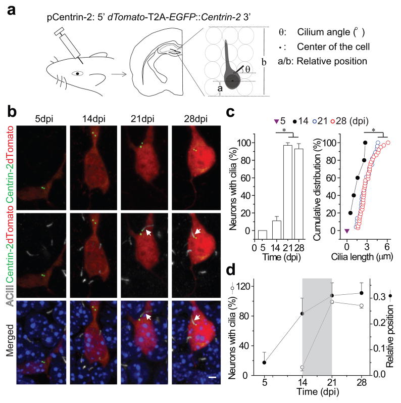
Figure 1. Primary cilia assemble in developing adult-born neurons.
(a) A schematic diagram of the retroviral vector (pCentrin-2), retroviral injection, a sample brain section and the parameters measured from reconstructed images. (b) Confocal images of cilia formation in developing adult-born neurons showing EGFP (Centrin-2), dTomato, DAPI and immunostaining for ACIII. Arrows point to primary cilia associated with centrosomes. Scale bar: 3 μm. (c) Quantification of percentage of labeled adult-born neurons with primary cilia (left) and the distribution of ciliary length (right) at 5, 14, 21 and 28 dpi. (d) Percentage of retrovirally labeled neurons with primary cilia and their relative position within the dentate gyrus granule layer at 5, 14, 21 and 28 dpi. Values represent mean±SEM (n=32–48 neurons; *: p<0.01, ANOVA or Kolmogorov-Smirnov test). The analyses in c and d are from the same group of cells.

