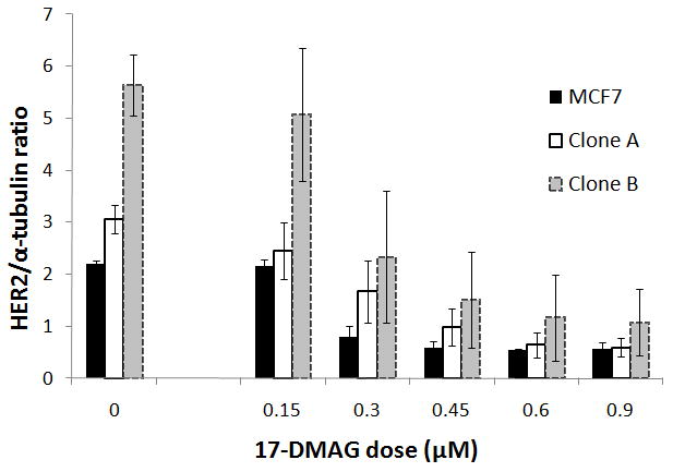
Figure 2.

Western blots confirmed low (MCF7 parental) intermediate (Clone A) and high (Clone B) Her2 expression. On the left (no 17-DMAG, dose 0), samples of 3 experiments were loaded into 1 gel to show the standard error of the mean. Her2 expression decreased dependent on the 17-DMAG dose added to the cells (incubation time was 24 hours). Data was normalized to the mean Her2 expression for each cell line at dose 0 in the same gel. All experiments were repeated 3 times. Results are statistically significant (p < 0.05) from 0.45 μM for Clone B and Clone A, and from 0.30 μM for MCF7 parental cells. Error bars represent the standard error of the mean.
