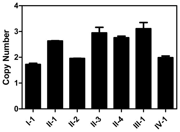
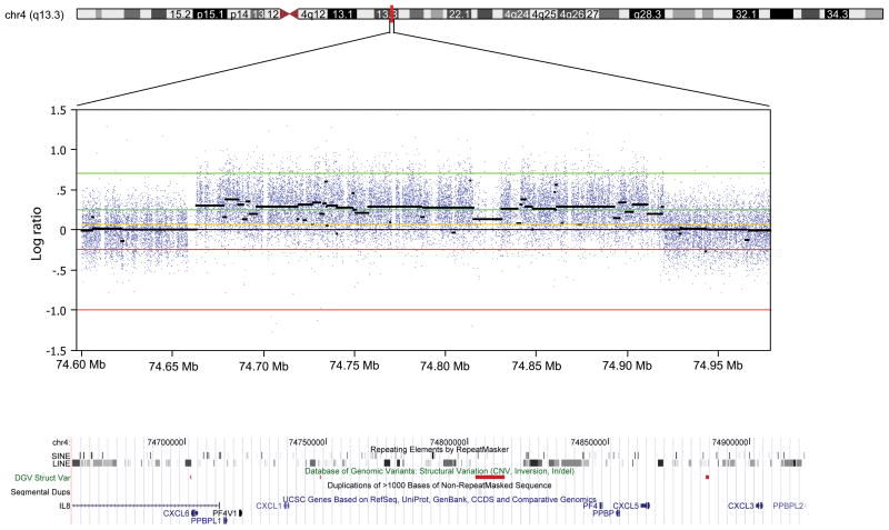
Fig. 2.
The 4q13 duplication identified in the melanoma-prone family. Panel a. Quantitative PCR (qPCR) of CXCL3 in genomic DNA of the melanoma family. Each qPCR assay was performed in duplicate. Results of qPCR assays for each individual are shown as a point estimate and 1 SD interval expressed as fold-difference compared to a reference sample. Data for CXCL6 were similar (not shown). Panel b. The duplication in melanoma patient II-3 by 4q13-focused array-CGH. Previously-reported CNVs listed in the Database of Genomic Variations are shown as red bars and none were located within genes. The duplicated region contains short and long interspersed repeat elements (SINE and LINE). Panel c. Breakpoint junction of the duplication showing a head-to-tail orientation. Tel-REF: telomeric reference sequence; Cen-REF: centromeric reference sequence. Matched sequences between reference and the melanoma patient are shown in colors (blue for telomeric and red for centromeric).


