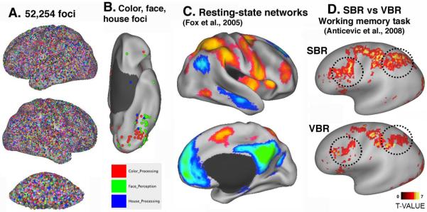
Figure 6. Multiple ways to map fMRI activations to PALS-B12 atlas surfaces in Caret.
A. 52,254 stereotaxic foci from 1,636 studies from the SumsDB Foci Library mapped to the inflated cerebral and cerebellar atlas surfaces. B. Foci specifically involved in processing of color (red), faces (green), or houses (blue) on a ventral view of the left hemisphere. C. Resting-state networks mapped to the atlas surface using average fiducial mapping (Fox et al., 2005). D. Working memory activations from 29 schizophrenia patients analyzed by Landmark-SBR (top) vs affine-registered VBR (adapted, with permission from Anticevic et al., 2008).

