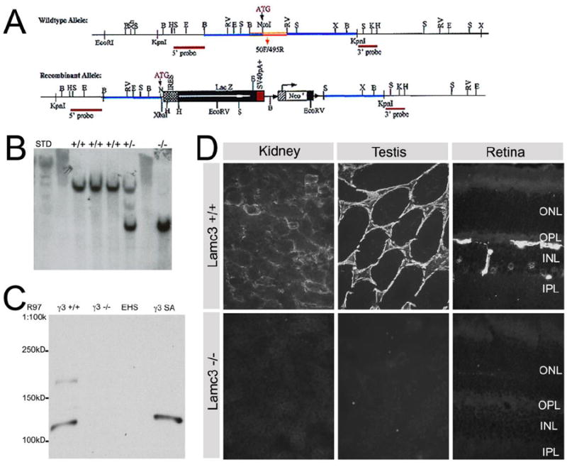
Figure 1.

Generation and verification of Lamc3-/- mice. A: A schematic view of Lamc3 wild type and recombinant alleles. The targeting construct was created by cloning a 3.6kb BamHI fragment containing the promoter and 5′ UTR of the Lamc3 gene together with the SalI fragment of pGT1.8 Ires βgeo (Mountford et al., 1994) and a 4.3kb EcoRV-KpnI fragment of intron 1 of Lamc3. The resulting targeting vector created a 2.2kb deletion spanning exon 1 and part of intron 1 of the Lamc3 gene and replaced with a promoter-less IRES β-Geo cassette. The neo cassette was subsequently removed. B: Southern blot screening of HindIII digests of genomic DNA for Lamc3+/+, Lamc3+/- and Lamc3-/- mice, probed with the “5’ probe”. The endogenous allele is present as a ~10.5 kb band; the recombinant allele is present as a ~5.9 kb band. C: Western blot of Lamc3+/+ (γ3+/+) and Lamc3-/- (γ3-/-) retina homogenates using the γ3 chain antiserum R97. EHS laminin (EHS) and recombinant γ3 chain short arm protein (γ3SA) were controls. R97 reacted with γ3SA at about 120kD. R97 also reacted with bands of 120kD and 180kD in the wild-type (γ3+/+). No reactivity was observed with Lamc3-/- homogenates (γ3-/-) or with EHS laminin. D: Immunohistological staining of Lamc3+/+ and Lamc3-/- kidney, testis and retina using anti-γ3 chain antibody R96. There was no detectable laminin γ3 IR in the Lamc3-/- tissues.
