
Figure 2.
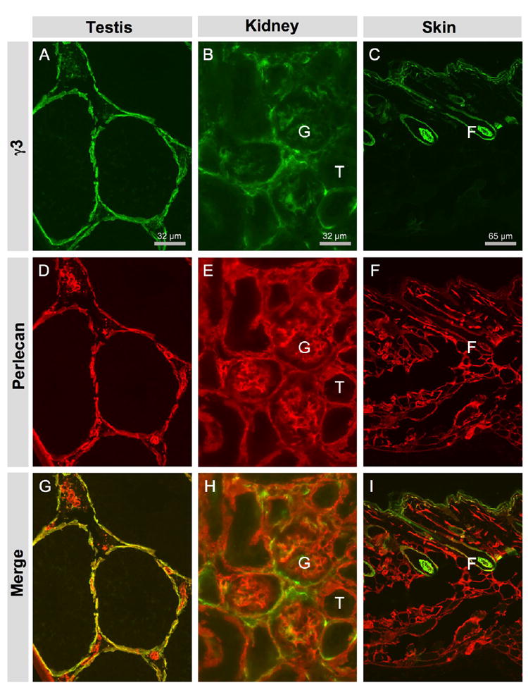
Laminin γ3 chain is widely but unevenly distributed in the basement membranes of various tissues. A, D, G: testis. B, E, H: kidney. C, F, I: skin. A, B, C: γ3 IR in testis, kidney and skin, respectively. D, E, F: Perlecan IR marks the BMs in these tissues. G, H, I: Overlay of γ3 IR and perlecan IR. A: In testis, γ3 IR is mainly located at the BM of seminiferous tubes; some γ3 IR is located at the BM of interstitial structures, presumably vasculature. D: In testis, perlecan IR is more broadly distributed. B: In kidney, γ3 IR is present mainly in glomerular (G) BM and somewhat weakly and discontinuously in the tubular (T) BM. E: In kidney, perlecan IR is evenly distributed in the BMs of kidney tubules and in the GBM. C: In skin, γ3 IR is located at the BM of the epidermis and around the hair follicle (F). F: In skin, perlecan is more widely distributed throughout the skin.