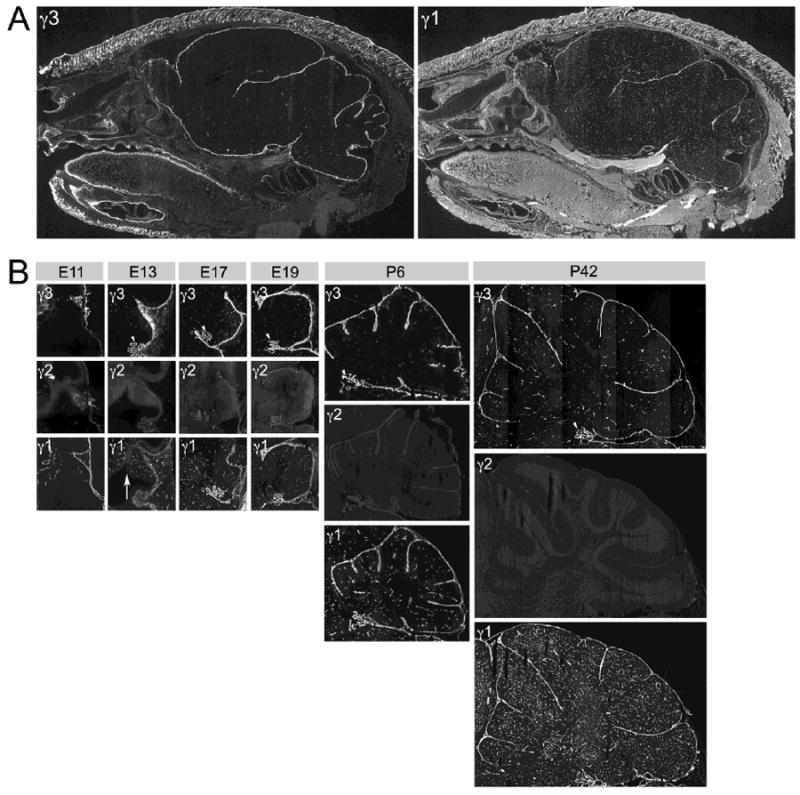
Figure 5.

Spatial and developmental expression of laminin γ chains in the brain. A: Sagittal sections of P6 mouse head. Adjacent sections reacted with laminin γ3 and γ1 antibodies, respectively. Laminin γ3 chain IR is present in various BMs of the brain, such as the pial and vascular BMs. Laminin γ1 chain IR is more widely distributed, not only in the CNS, particularly in the vasculature, but also in other tissues (e.g., the tongue). B: Developmental expression of all three laminin γ chains in the mouse cerebellum, from E11 through P42. Laminin γ3 chain IR is present in various BMs during development. Laminin γ2 chain IR is nearly absent at all ages. Laminin γ1 chain IR is most widely distributed at all ages. Arrowheads mark the choroid plexus; the arrow marks the ventricular zone. See text for details.
