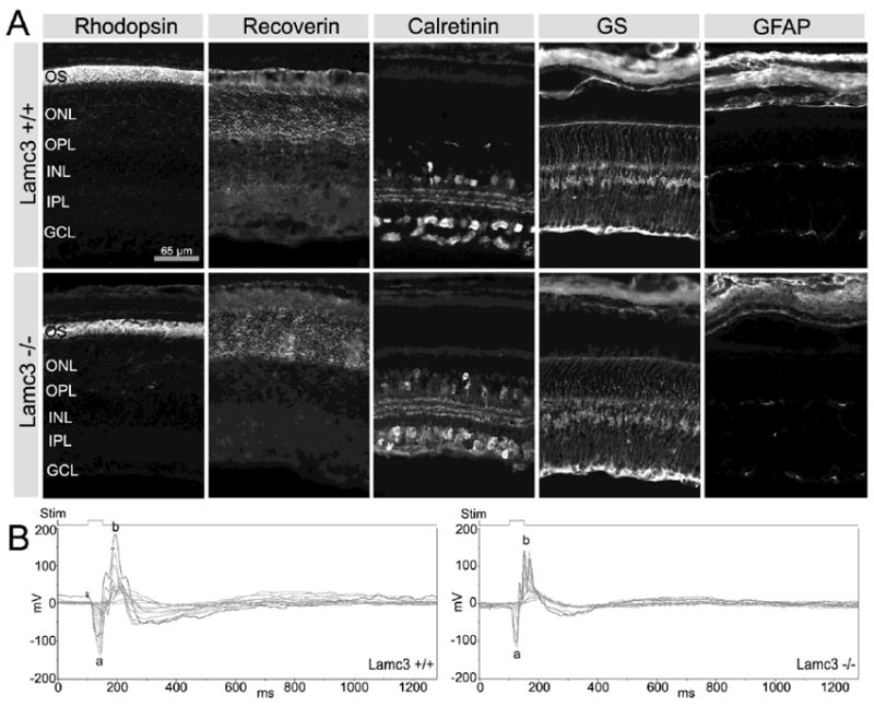
Figure 8.

Comparison of cell markers and ERGs in Lamc3+/+ and Lamc3-/- retinas. A: Photoreceptor outer segment (OS) labeled with anti-rhodopsin, photoreceptors labeled with anti-recoverin, amacrine cells labeled with anti-calretinin, Müller cells labeled with anti-glutamine synthetase (GS), and astrocytes labeled with anti-GFAP display no obvious differences between the Lamc3+/+ and Lamc3-/- retinas. B: The Lamc3-/- ERG has both a- and b-waves, although the amplitude of the Lamc3-/- b-wave is smaller than that in the Lamc3+/+. Thin lines, ERG traces of each trial; thick lines, the average of individual trials. Light flash (Stim), 50 ms.
