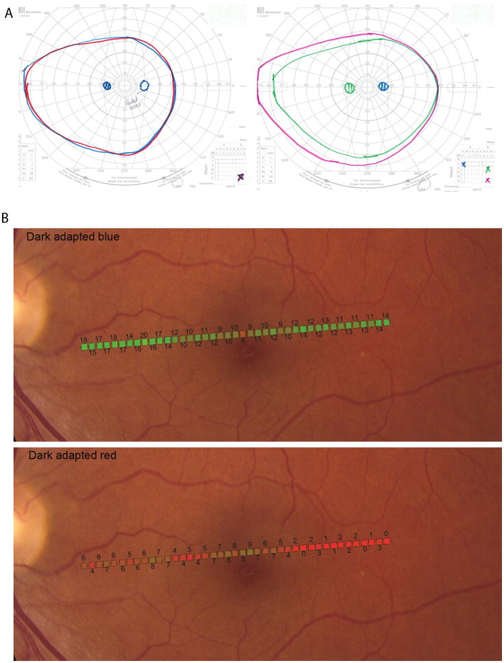
Figure 3. Acute Zonal Occult Outer Retinopathy.
(Top) Kinetic perimetry for patient 2, left eye. (Left) Two-color dark-adapted Goldmann kinetic field isopters. The isopter for the V3c long-wavelength stimulus target (red solid line) and the isopter for the V3̅c short- wavelength stimulus target (blue solid line) are superimposed. The isopter for the II3c long- wavelength stimulus target and the isopter for the II3̅c short- wavelength stimulus target (not shown) were also superimposed. In these rod-mediated tests, the scotoma is absent and both red and blue stimuli were seen as achromatic, although the patient reported the blue targets to appear “blurry or dimmer” in the region of the scotoma. (Right) Light-adapted, Goldmann kinetic field isopters obtained by using a white target stimulus shows full peripheral fields with the II3c and V3c targets; the I4e stimulus shows the scotoma.
(Bottom) Dark-adapted two-color fundus-guided microperimetry for patient 2, left eye. The data from microperimetry is superimposed onto a fundus image. (Top) Dark adapted blue (Blue DA) sensitivities were normal throughout the regions tested (background illuminance of 0cd/m2, 200 ms Goldmann V blue stimulus, 2.0 neutral density (ND) filter. (Bottom) Dark adapted red (Red DA) sensitivities showed a dense scotoma beginning about 3 degrees temporal to fixation (background illuminance of 0cd/m2, 200 ms Goldmann V red stimulus, 1.0 ND filter). The difference between Blue DA and Red DA sensitivity values for each retinal location in the region of scotoma was at least 8dB, demonstrating rod-mediated sensitivity in the scotomatous region.

