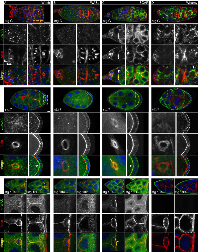
Fig. 4.
WASP family protein co-localization with F-actin during Drosophila oogenesis. A-D: Confocal micrograph projections showing WASP family proteins (green or grayscale) co-localizing with F-actin (red or grayscale) and co-stained with DAPI (blue) as indicated. Wash (A), WASp (B), SCAR (C) and Whamy (D) expression patterns in the germarium (upper panels) and individual egg chambers from stages 7 (middle panels) or stages 10A and 10B (lower panels) showing co-localization with actin rich structures. Higher magnification views of germarium region 1 (yellow dashed box) and regions 2b-3 (red dashed box), stage 7 ring canals (red dashed box) and oocyte cortex (yellow dashed box), stage 10A border cells (red dashed box) and nurse cell actin bundles in stage 10B (yellow dashed box). Scale bars: Germarium: 10µm, stages 7, 10A and 10B: 100µm.

