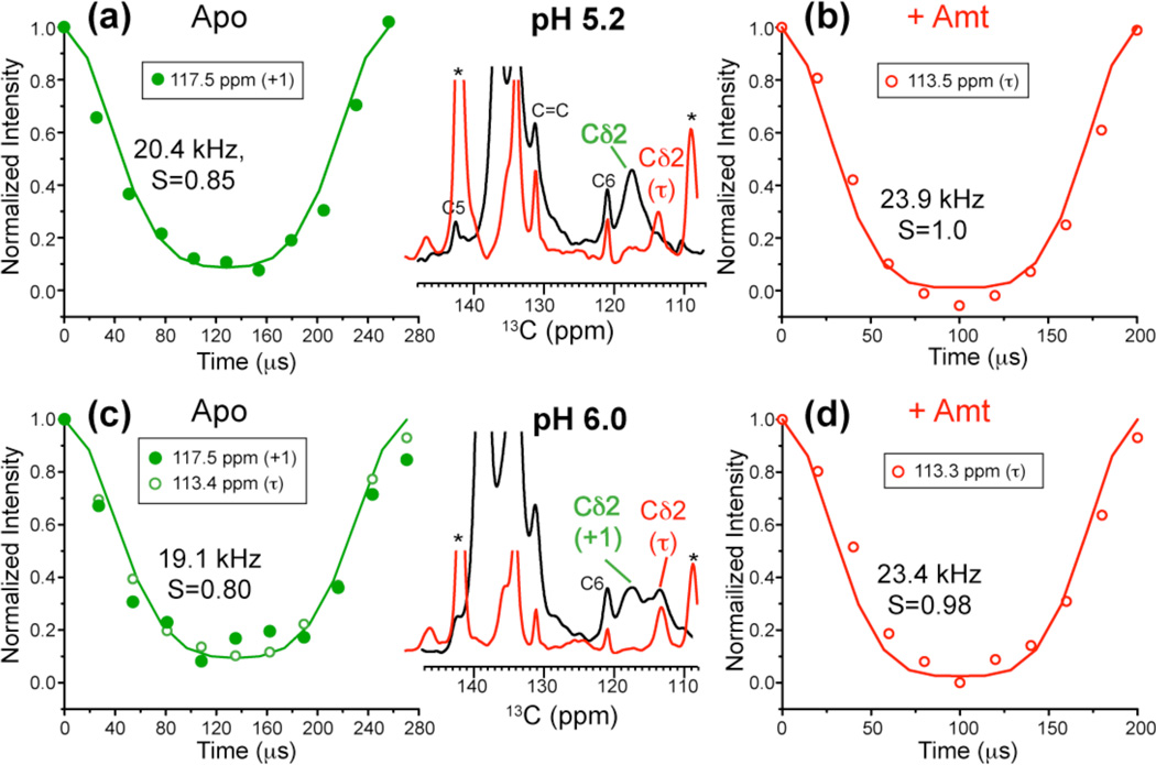
Fig. 8.
His37 sidechain dynamics at mildly acidic pH and 308 K from Cδ2-H dipolar couplings. (a) Apo peptide at pH 5.2. (b) Amt-bound peptide at pH 5.2. (c) Apo peptide at pH 6. (d) Amt-bound peptide at pH 6. The aromatic regions of the 13C spectra of the drug-free (black) and drug-bound (red) samples are compared in the middle column. The data were measured under 3.9 and 3.7 kHz MAS for (a, c) and under 5 kHz MAS for (b, d). The best-fit C–H dipolar coupling value and the corresponding bond order parameter are indicated for each curve.

