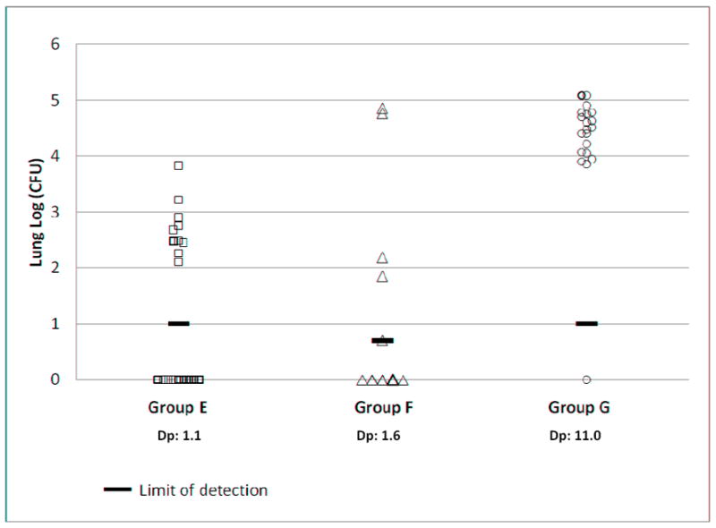
Figure 2. MTB CFU in lungs after ULD exposure.

Aerosol exposure groups are summarized in Table 2. CFU was determined by lung necropsy of groups E, F, and G at 5, 3, and 4 weeks, respectively post aerosol exposure. The limit of detection (LOD) is shown for each group. When no bacteria were grown, the results are plotted on the X-axis. Dp: presented dose (CFU/mouse).
