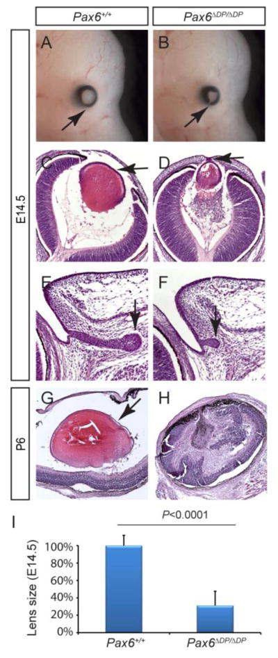
Figure 4. Defective lens and lacrimal gland development in Pax6ΔDP/ΔDP mutants.

(AB) The E14.5 Pax6ΔDP/ΔDP mutant exhibited coloboma, due to a failure of optic cup closure (arrows). (C–D) Compared to the wild type, the Pax6ΔDP/ΔDP mutant lens was much reduced in size and remained attached to the surface ectoderm (arrows). (E–F) The lacrimal gland budding was stunted in Pax6ΔDP/ΔDP mutant. (G–H) By P6, the lens completely degenerated in Pax6ΔDP/ΔDP mutant. (I) Quantification of lens size at E14.5. [Student’s t test: P<0.0001 for the Pax6ΔDP/ΔDP mutants (n=4) compared to the wild type (n=6).]
