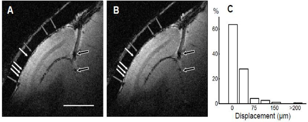
Figure 4. Reproducibility of structural images within a session.
(A and B) Structural images collected in two runs (30 minutes each) that were one hour apart (slice thickness, 1 mm; in-plane resolution, 100 × 100 µm2). The locations of the pial veins (black and gray arrows) and principal veins within gray matter (white arrows) are the same. Scale bar: 5 mm. (C) The distribution of relative displacement between runs in the duration of 30 minutes (the length of a typical run).

