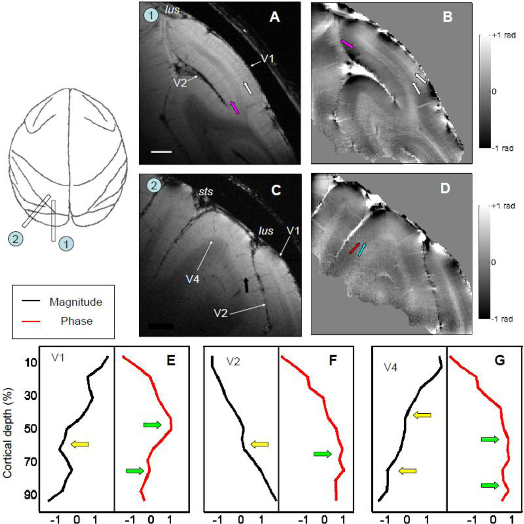
Figure 8. MR structural images of extrastriate visual cortex.
The magnitude (A) and phase (B) images of a parasagittal slice revealed V2 buried in the lunate sulcus (lus). Besides the stripe of Gennari (white arrow) in V1, a dark layer (pink arrow) can be found in the middle of V2 from the magnitude map (A). From the phase map (B), one bright layer exists in V2 (pink arrow) in additional to the two bright layers in V1 (white arrows). (C) and (D) An oblique slice that includes the portion of V4 that located between superior temporal sulcus (sts) and lus. Laminar structures within V4 (black arrows) can be found both from magnitude map (C) and phase map (D). The averaged results of magnitude (black lines) and phase profiles (red lines) from prestriate visual cortex of V1 (E), V2 (F), and V4 (G) are summarized. The peaks in phase profiles and dips in magnitude profiles are marked by green and yellow arrows, respectively. Both images were acquired with in-plane voxel size of 100 × 100 µm2, thickness of 1 mm, and flip angle of 45°. Scale bar: 2 mm in (A).

