
Figure 1.
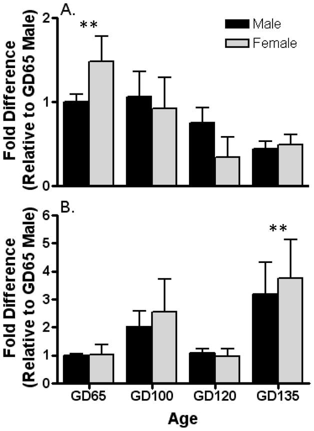
Developmental changes in expression of aromatase mRNA in the left medial preoptic area-anterior hypothalamus (a) and amygdala (b) of fetal sheep. Expression of aromatase mRNA is normalized to GAPDH mRNA expression in the same sample. Data are the mean ± SE fold differences relative to mean expression of 65-day-old male fetuses (n = 3–6 animals per group). Analysis of the data by 2-way ANOVA revealed a significant age effect (P < 0.05), but no sex effect or interaction in both the medial preoptic area and amygdala. Thus, the data for the sexes were combined and reanalyzed by a Kruskal-Wallis 1-way ANOVA followed by Dunn’s multiple comparison test. Poshoc analysis revealed that GD 135 fetuses differed from other ages by exhibiting significantly lower aromatase mRNA expression in the medial preoptic area and significantly higher expression in the amygdala (**, P < 0.05 vs. all other ages).