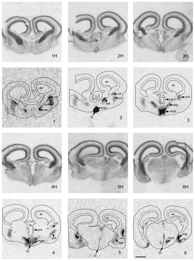
Figure 2.
Images of coronal sections through the preoptic area, basal hypothalamus and amygdala of a GD 53 lamb fetus showing the expression of aromatase mRNA. Images are arranged in a rostral to caudal order. 1H–6H are the hemotoxylin stained images, that match the autoradiographic images (1–6) of aromatase mRNA expression generated by in situ hybridization using 33P-labeled cRNA probes. The following anatomic sites of aromatase mRNA expression were distinguished: avpv, anteroventral periventricular preoptic area; bst, bed n. of the strial terminalis; ca, cortical amygdala; cd, caudate n.; ma, medial amygdala; mpa, medial preoptic area; *, nascent ovine sexually dimorphic n.; nac, n. acumbens; put, putamen; pvt, periventricular thalamic area; vmn, ventromedial n. of the hypothalamus. Other abbreviations: f, fornix; 3v, 3rd ventricle; 4v, 4th ventricle. Scale bar measures 2 mm. Brightness and contrast were adjusted to optimize image.

