
Fig. 6.
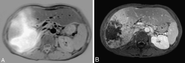
Pre- and post-Yttrium-90 radioembolization. a: Fusion image of liver MRI and 99mTc-MAA scintigram showing large HCC lesion in right liver lobe. b: Contrast-enhanced MRI 3 months post-treatment shows necrotic zone centrally in HCC
Official websites use .gov
A
.gov website belongs to an official
government organization in the United States.
Secure .gov websites use HTTPS
A lock (
) or https:// means you've safely
connected to the .gov website. Share sensitive
information only on official, secure websites.
Pre- and post-Yttrium-90 radioembolization. a: Fusion image of liver MRI and 99mTc-MAA scintigram showing large HCC lesion in right liver lobe. b: Contrast-enhanced MRI 3 months post-treatment shows necrotic zone centrally in HCC