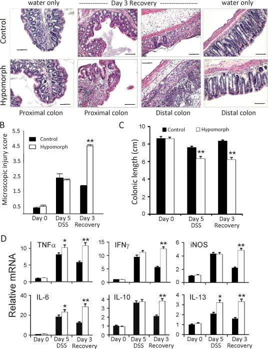
Figure 3.
Intestinal tissue recovery is impaired in St14 hypomorphic mice. (A) Representative H&E stained sections of colonic tissue segments from St14 hypomorphic and littermate control mice after 7 days of DSS and 3 days water (Day 3 Recovery), showing the persistence of inflammatory infiltrates in the proximal and distal colons of St14 hypomorphic mice compared with the well-defined colonic crypts, and a normal submucosa layer of water alone-treated mice. Scale Bars, 100μM. (B) Comparison of microscopic injury in the proximal colons of St14 hypomorphic and littermate control mice on the indicated days, showing persistent injury in St14 hypomorphic mice. n=4-5; **p <0.005, Mann Whitney U Test. (C) Colonic lengths measured during the acute and recovery phases of DSS induced injury. n=7; **p <0.005, Mann Whitney U Test. (D) Comparison of relative mRNA levels of cytokines and other mediators in distal colons of St14 hypomorph (open bars) and littermate control mice (solid bars) during the DSS protocol, measured by qPCR analyses. n = 3-4 mice per group. *p <0.05, **p <0.005, unpaired Student t test.

