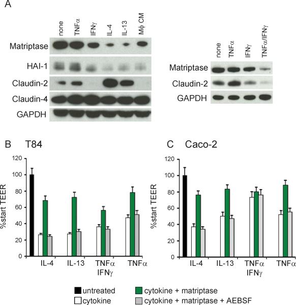
Figure 7.
Loss of matriptase contributes to intestinal epithelial barrier disruption by inflammatory cytokines. (A) Polarized T84 monolayers on transwell filters were treated daily on the basolateral side with 10ng/ml TNFα, IL-4, or IL-13, 100U/ml IFNγ, a combination of TNFα and IFNγ, 10% macrophage (Mϕ) conditioned media or left untreated. Protein lysates were analyzed at 48hrs by immunoblotting for matriptase, HAI-1, claudin-2, claudin-4, and GAPDH. (B) Polarized T84 or (C) Caco-2 monolayers were treated for 48hrs with the indicated cytokines as in (A), then replaced with 5nM recombinant matriptase or media alone control. TEER was measured after 8 hrs.

