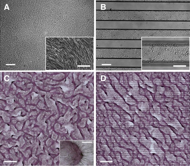
Figure 1. LR asymmetry in pattern formation by vascular mesenchymal cells.
Phase contrast microscopy of VMCs on (A) conventional plastic substrate at confluence and (B) FN/PEG substrates (interfaces identified by black titanium lines) showing preferential attachment to FN domain immediately after plating. Scale bar, 300 μm and 200 μm (inset). After 10-14 days, development of regularly spaced aggregates (C) in a labyrinthine configuration and (D) in a stripe pattern along principal diagonal axis in bright field (multicellular ridges stained with hematoxylin). Insets: higher magnification images of multicellular aggregates. Scale bar, 2 mm and 300 μm (inset).

