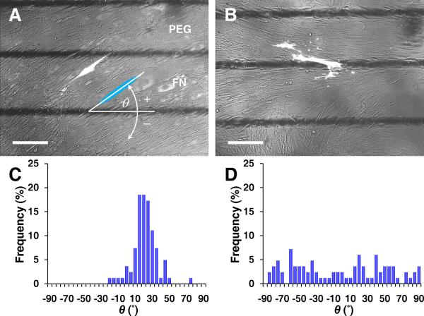
Figure 2. Early stage of coherent single-cell orientation perpendicular to the axis of diagonal ridges.
Fluorescence microscopy of GFP-transfected VMCs on (A) FN/PEG substrates showing coherent individual cell orientation, relative to the interface axis (black lines) and (B) control substrate as homogeneous FN. Scale bar, 200 μm. Histogram of θ for (C) FN/PEG, showing convergence to 19 ± 14° (n = 81 cells; day 5; mean ± s.d.) and (D) control substrate, showing non-convergence (n = 83 cells; day 5).

