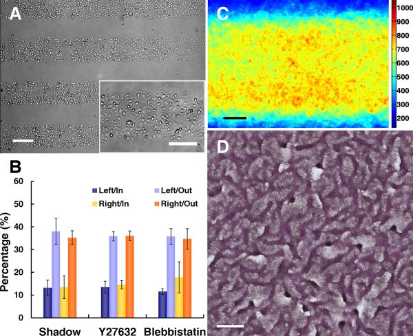
Figure 7. Abrogation of LR polarity and LR alignment of multicellular ridges with removal of substrate interfaces.
A, Microscopic images after shadow-mask plating show plating limited to rectangular domains. Scale bar, 300 μm and 200 μm (inset). B, Polarity of VMCs at the edges of cell sheets shadow-plated onto uniform FN (n = 3 experiments; > 175 cells each; mean ± s.d.) and VMCs near the FN/PEG interface with Y27632 (n = 3; > 135 cells each) or blebbistatin (n = 3; > 150 cells each) inhibition. C, Stacked images of NMM-IIa immunofluorescence after shadow-plating on FN (n = 40). Scale bar, 100 μm. D, Multicellular patterns by hematoxylin staining after shadow-plating on FN substrate. Scale bar, 2 mm.

