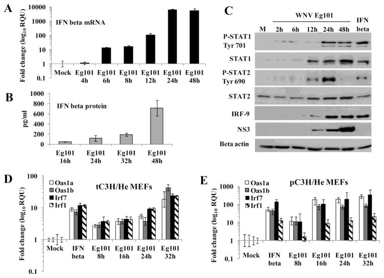
Figure 1. IFN beta is produced by WNV Eg101 infected MEFs and induces phosphorylation of STAT1 and STAT2.
MEFs were mock-infected or infected with WNV Eg101 at a MOI of 5 for the indicated times or treated with 1000 U/ml of murine IFN beta for 3h. (A) Total RNA from tC3H/He MEFs was extracted and IFN beta mRNA levels were measured by real-time qRT-PCR. (B) Extracellular IFN beta protein levels in tC3H/He MEF culture fluid was measured by ELISA. The values shown are averages from at least two independent experiments performed in triplicate. The error bars represent SDM. (C) tC3H/He cell lysates were analyzed by Western blotting using antibodies specific for the indicated proteins. Actin was used as the loading control. The blots shown are representative of results obtained from three independent experiments. (D) and (E) Comparison of ISG activation by IFN beta in primary and transformed MEFs. Total RNA was extracted from (D) tC3H/He or (E) pC3H/He MEFs infected with WNV Eg101 (MOI 5) for the indicated times, mock-infected or treated with murine IFN beta (1000 U/ml) for 3h. The changes in the levels of Oas1a and Oas1b mRNA were assessed by real-time qRT-PCR. Each experiment was repeated at least two times in triplicate. The mRNA level of each gene was normalized to the level of GAPDH mRNA in that sample and is shown as the fold change over the amount of mRNA in mock samples expressed in relative quantification units (RQU). The error bars represent the calculated SEM (n = 3) and are based on an RQMin/Max of the 95% confidence level.

