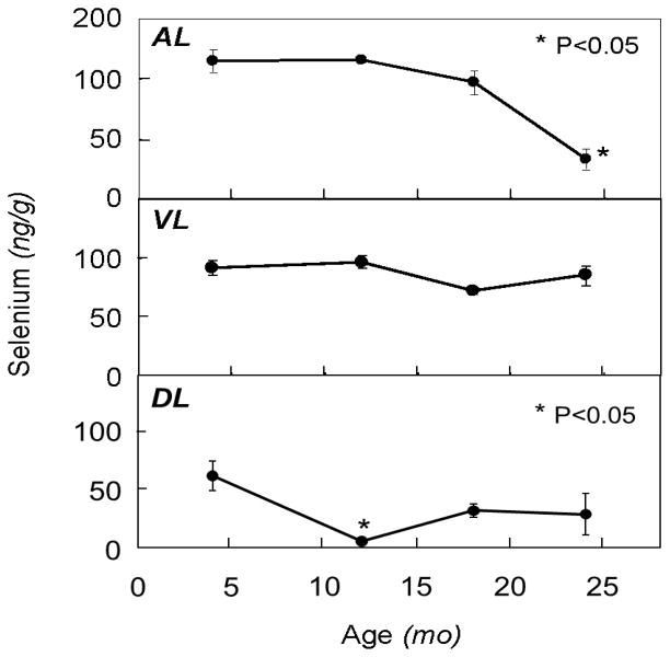
Figure 1.
Effect of aging on levels of selenium in different prostate lobes of the rat. Rats were sacrificed at 4, 12, 18 and 24 months of age and prostate lobes (AL, anterior; VL, ventral; DL, dorsolateral) were removed and analyzed for selenium content as described in text. Points and error bars represent mean ± SEM (n=3–6). Asterisks reflect statistical significance compared to the young group (P<0.05).

