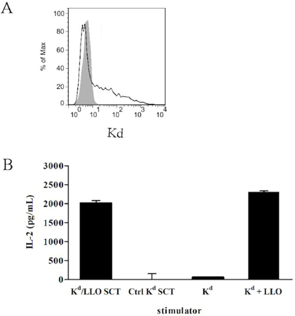
Figure 1. Expression and recognition of Kd/LLO91–99 SCTs by CD8+ T cells.
(A) Cell surface expression of Kd/LLO91–99 SCTs. 293T cells were transfected with Kd/LLO91–99 SCT DNA and stained with anti-Kd mAb (SF1-1.1.1, solid line). A shaded histogram shows staining of untransfected cells. (B) Activation of hybridomas specific for LLO 91–99/Kd by Kd/LLO91–99 SCT-expressing cells. Transfected 293T cells were incubated with T hybridomas at the ratios of 1:1 for 24 hours and the amounts of IL-2 in the culture supernatant were measured by ELISA. For controls, P815 cells that express endogenous Kd were incubated with hybridomas with or without 1µg/mL of LLO 91–99 peptide.

