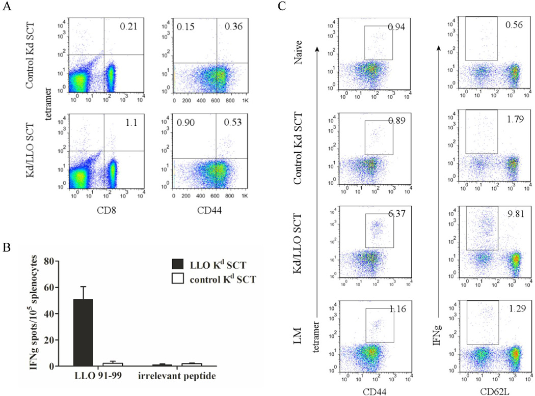
Figure 3. A SCT DNA vaccine develops memory CD8+ T cells.
Mice were vaccinated with Kd/LLO91–99 SCTs or control Kd SCTs as described above and rested for 5 months before L. monocytogenes challenge. (A) At 5 months post-immunization, splenocytes were stained with anti-CD8, -CD44 mAb and Kd/LLO91–99 tetramers. In the left panels CD3 positive cells are shown and the numbers indicate the percentage of CD3+ CD8+ tetramer+ cells. In the right panels CD3+ CD8+ cells are shown and the numbers indicate the percentage of cells in each quadrant. The flow cytometry data are presented from one representative experiment of three performed independently. (B) Splenocytes from each group were stimulated in vitro with LLO 91–99 or irrelevant peptide overnight and IFNγ secretion was measured by ELISPOT. Error bars indicate SE of the experiment. The data presented are from one representative experiment of two performed independently with each group containing 4~5 mice. (C) Vaccinated mice were challenged with 103 L. monocytogenes. At 7 days post-infection, splenocytes were stained with anti-CD8, -CD44 mAb, and Kd/LLO91–99 tetramers (left) or with anti-CD8, CD62L and -IFNγ mAb after 4 hour stimulation in vitro with LLO or irrelevant peptide in the presence of Golgi-blocking agent (right). CD8 positive cells were shown. Numbers indicate the percentage of cells of the gated population among CD8+ cells. Representative figures of the flow cytometry data.

