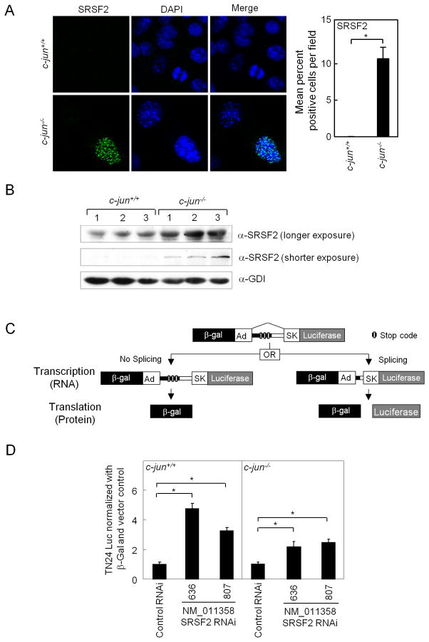
Figure 5. Endogenous c-jun determines the relative abundance of the pro-apoptotic splicing factor SRSF2.
(A) Immuno-fluorescence staining for SRSF2 (green nuclear speckles) and nuclear stain (DAPI: blue) was performed on MECs from c-jun+/+ and c-jun−/− and quantification of percentage positivity within 5–10 microscopic fields (B) Western blotting of the SRSF2 in c-jun+/+ and c-jun−/− KO MECs. (C) Schematic representation of the double reporter splicing reporter with two transgenes, the β-galactosidase (β-gal) and luciferase (Luc). Reporter genes are fused in frame by recombinant fragments of the genes encoding adenovirus (Ad). The recombinant fragment contains skeletal muscle isoform (SK) of human tropomyosin. Three in-frame translation stop signals (XXX) are present in the intronic region. (D) Relative splicing activity measured as relative luciferase activity divided by total β-galactosidase (luciferase units × 103) is shown comparing c-jun+/+ and c-jun−/−. Cells were treated either with two different SRSF2 siRNA, or control siRNA. Activity of c-jun−/− and c-jun+/+ were equalized to 1 in the presence of control siRNA to illustrate the effect of SRSF2 siRNA. The assay was conducted in fibroblasts (MEFs) or mammary epithelial cells (MECs) generated from floxed c-jun transgenic mice (c-jun+/+ or c-jun−/−). The data represents mean ± SEM of n>5 separate transfections. *p ≤ 0.05, EB= SEM.

