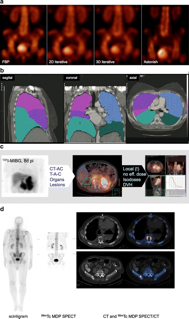
Fig. 7a–d.

Examples of methodological advances in SPECT/CT imaging and future applications. a Advanced three-dimensional (3D) iterative SPECT reconstruction (Astonish, Philips Healthcare) incorporating projection-based collimator/COR distance information, collimator resolution, noise reduction and attenuation/scatter correction. (Courtesy of Antonis Kalemis, Philips Healthcare) b An example of a segmented CT of the thorax showing the individual lobes of the lung. The colours indicate different regions of interest, which are applied to the co-registered lung image to derive anatomically accurate functional values for ventilation and perfusion from the SPECT lung scan. (Courtesy Dale Bailey, Royal Northshore Hospital, Sydney, Australia) c Voxel-based dosimetry from 3D SPECT(/CT). SPECT data following CT-AC are collected for multiple time points. CT-based organ segmentation and SPECT-based lesion segmentation is performed on SPECT/CT images. Local dosimetry is possible and isodose curved and dose-volume histograms can be calculated. (Courtesy of Bernd Schweizer, Philips Healthcare) d Whole-body SPECT(/CT): patient with severe back pain with multiple focal uptakes on the scintigram. A contiguous two-bed SPECT/CT acquisition reveals several bone metastases in the spine and pelvis. Multi-bed, or whole-body SPECT/CT helps with the complete restaging of the patient, and is in synchrony with similar referring indications for whole-body PET/CT, which is already widely available. (Courtesy of Ora Israel, Haifa)
