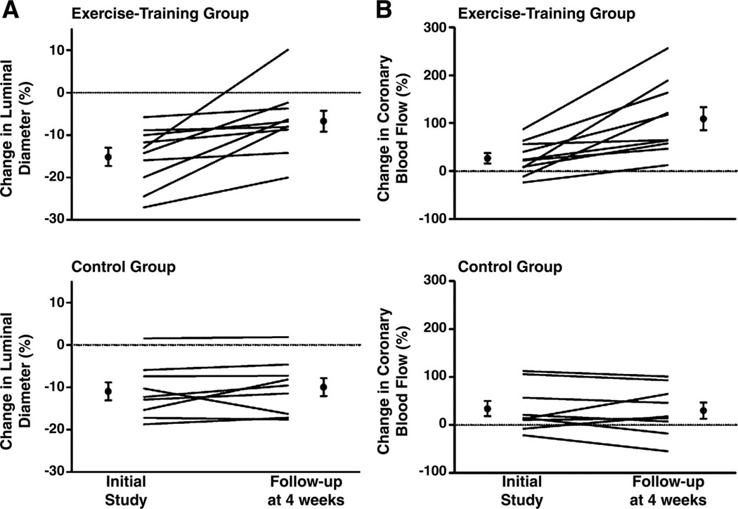
Fig. 2.
Changes in coronary artery luminal diameter (A) and coronary blood flow (B) in response to intracoronary administration of acetylcholine in exercise-trained (top) and sedentary control (bottom) subjects. Lines show changes in individual subjects and black dots show mean ± SE for the groups. Data are taken with permission from Hambrecht et al. (49) ©[2000] Massachusetts Medical Society. All rights reserved.

