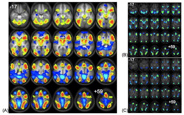
Figure 1.
Left Panel (A): Statistical map showing group composite activations in the “generate” (hot colors) versus the “read” condition (cool colors). Statistics for the clusters are listed in Table 1A. Top Right Panel (B): Statistical map showing group composite activations in the “generate” relative to the “read” condition in older adults ages 35–62. Bottom Right Panel (C): Statistical map showing group composite activations in the “generate” relative to the “read” condition in younger adults ages 19–34. All images are in radiological convention (right on the image corresponds to left in the brain), and span z-coordinates −17 to +59 in 4mm increments.

