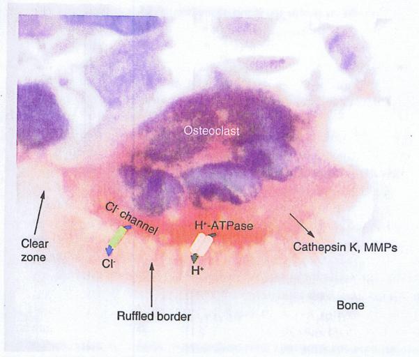
Figure 1. Osteoclast resorbing bone.
The ruffled border formed on the underside of the osteoclast facing the bone increases the surface area of the cell. The clear zone surrounds the ruffled border and seals the resorbing compartment. Hydrochloric acid is formed in the cavity following active transfer of H+ by a proton pump (the vacuolar-type H+-ATPase) and passage of Cl− via a Cl− channel, and proteolytic enzymes are released passively from the osteoclast into the cavity to dissolve the mineral and matrix components of bone, respectively. (Section of calvarial bone from a mouse treated with IL-1 and stained for tartrate-resistant acid phosphatase activity with hematoxylin counterstaining).
MMP: Matrix metalloproteinase.

