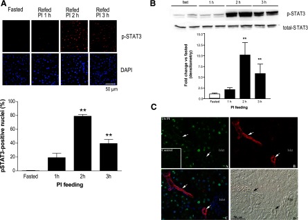
Fig. 5.
STAT3 is activated early in the course of cholecystokinin (CCK)-mediated growth of the exocrine compartment of the pancreas. A: immunohistochemistry for P-Tyr705 STAT3 and 4′6-diamidino-2-phenylindole (DAPI) nuclear stain in fasted mice or mice fasted and then refed PI-containing chow for 1, 2, or 3 h. Representative ×40 field (top) and quantitation of percent nuclear localization of P-STAT3 (bottom), with 3 mice per group (n = 3, **P < 0.01). B: representative Western blot (top) with summary of densitometry data below of p-Tyr705 STAT3 and total STAT3; 4 independent experiments (n = 4, **P < 0.01). C: immunohistochemistry for p-Tyr 705 STAT3 in exocrine pancreas of fasted (inset) and 2 h PI-fed mice (top left, A); immunohistochemistry for a ductal marker CK19 with arrows used for emphasis of duct-specific labeling (top right, B); overlay of p-Tyr705 STAT3 (green), ductal marker CK19 (red), and nuclear label DAPI (blue), showing the activation and nuclear localization of p-STAT3 occurs specifically in acinar cells, but not in islets or ducts (bottom left, C); corresponding Nomarski image (bottom right, D).

