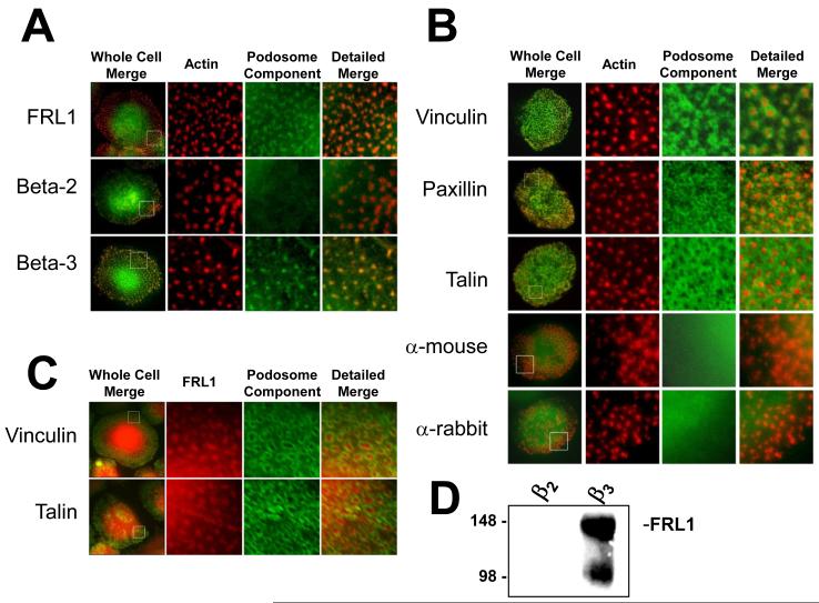
Figure 2. FRL1 Localizes to Podosomes in Differentiated Macrophages.
A. Fluorescence microscopy images of macrophages stained for FRL1 and integrins. The boxed areas in the first column are enlarged in the second, third and fourth columns, with the second column showing only rhodamine phalloidin staining of actin, the third showing only FITC staining, and the fourth showing a merged image of the second and third columns. FRL1 is visualized using a rabbit primary antibody followed by an FITC-tagged anti-rabbit secondary antibody. Integrins beta-2 and beta-3 are visualized using monoclonal mouse primary antibody followed by FITC-tagged anti-mouse secondary antibody. Boxed regions are 10μm by 10μm. B. The first column in each row shows a representative macrophage stained with rhodamine phalloidin and either no primary antibody, hVin α-vinculin, 349 α-paxillin, or 8D4 α-talin followed by FITC tagged anti-mouse or FITC tagged anti-rabbit. The boxed areas in the first column are enlarged in the second, third and fourth columns, with the second column showing only rhodamine phalloidin staining of actin, the third showing only FITC staining, and the fourth showing both. The anti-mouse and anti-rabbit controls show normal podosomes and demonstrate that the rhodamine phalloidin stain does not bleed into the green channel. Vinculin staining forms a concentric ring around the actin-rich podosome and is excluded from the core. Paxillin staining appears to surround the podosome and is excluded from the core but appears as punctute points surrounding the core rather than a continuous ring. Talin staining forms a modified sheet throughout the cell but is excluded from the actin-rich core. Boxed regions are 10μm by 10μm. C. The first column in each row shows a representative macrophage stained with goat anti-FRL1 antibody followed by TRITC-tagged anti-goat antibody and either 349 α-paxillin or 8D4 α-talin followed by FITC tagged anti-mouse or FITC tagged anti-rabbit. The boxed areas in the first column are enlarged in the second, third and fourth columns, with the second column showing only FRL1 staining, the third showing only FITC staining, and the fourth showing a merged image of the second and third columns. Boxed regions are 10μm by 10μm. D. Western blot of proteins immunoprecipitated from differentiated primary macrophage lysates using anti-β2 IB4 monoclonal antibody and anti-β3 7G2 and 1A2 monoclonal antibodies as described in Methods. IPs were separated by SDS-PAGE and blotted for FRL1. FRL1 co-immunoprecipitates with β3 integrin but not with β2 integrin.

