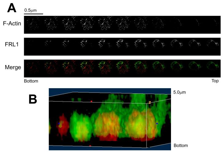
Figure 3. FRL1 Localizes above the Podosome Tip.
A. Z-axis kymograph composed of confocal microscopy images of a primary macrophage. Cells were fixed, permeabilized, and stained for FRL1 and actin using goat anti-FRL1 followed by FITC tagged anti-goat secondary antibody and rhodamine phalloidin, respectively. Progressive sections are incremented by 0.5μm. B. Three-dimensional reconstruction of podosomes stained for FRL1 and actin. FRL1 has been stained green using anti-FRL1 antibody followed by FITC-tagged secondary antibody while actin has been stained red with rhodamine phalloidin.

